

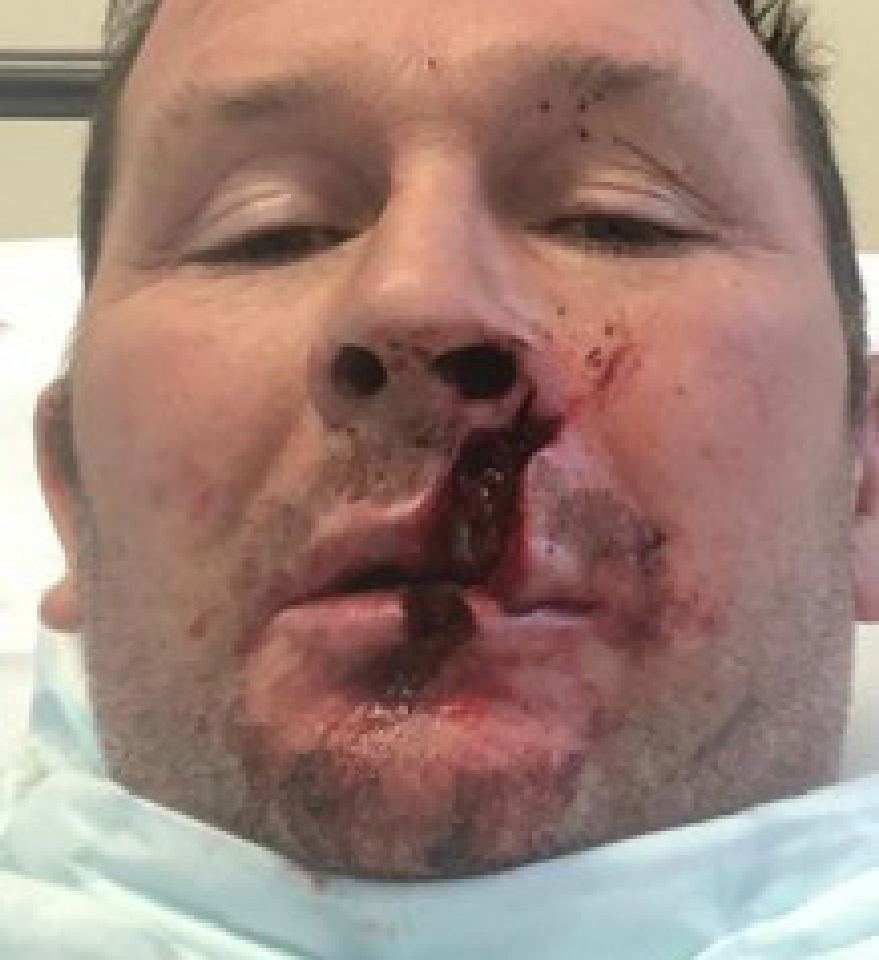
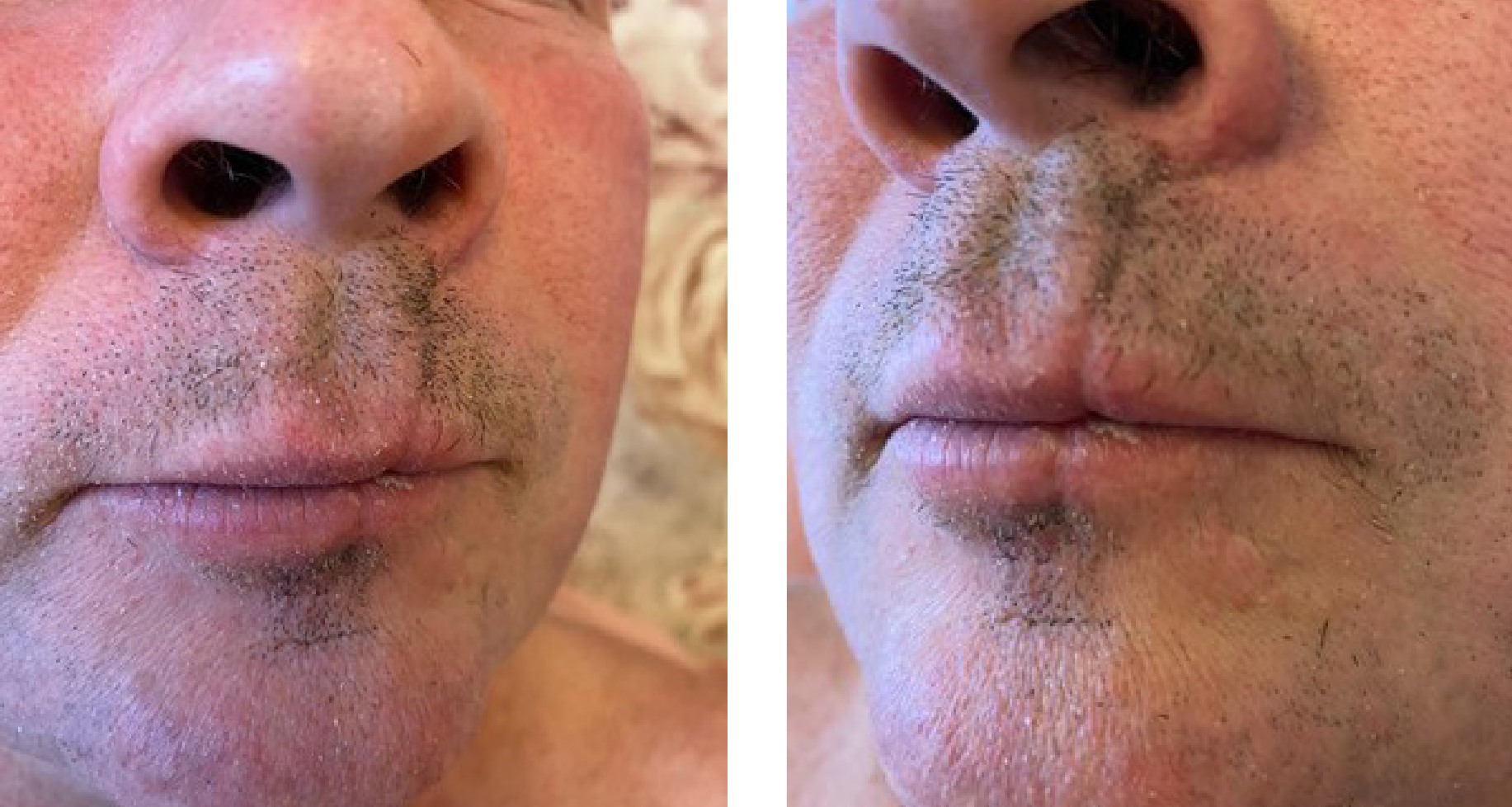
On 9 January 2021, a 50-year-old male (patient one) was injured by an angle grinder blade that was used without a safety guard. The blade kicked back and caused a facial injury to the left side of his face and across the lip. The patient was managed and treated in the accident and emergency department by a specialist maxillofacial team with suturing (Figure 1), as no other structures were involved, and he was discharged home the same day with no follow-up arranged. There was no other past medical history of note.
Management
A Silvery Blue light-emitting diode (LED) device and microneedling protocol was used to treat the patient.
The LED device is free-standing with five diode array panels with three different wavelengths that can be used solely or simultaneously with a variety of settings and treatment protocols. It is primarily used in the clinic post-treatment to aid healing and calm inflammation. The device was used here in a specially designed protocol. The wavelengths available are:
- For acne and inflammations: 415nm
- For wounds, burns, skin repair or post inflammation (i.e. laser, peels, microneedling): 635nm
- For deeper pain relief than 635nm: 830nm.
The general rule of thumb with light/laser therapy is that the further down the wavelength spectrum travelled, the deeper the penetration in the skin.
The patient attended the author's clinic on 19 January 2021 for his first in-clinic session (10 days post-accident) and, having consulted our clinical manufacturing partners regarding the best course of treatment, it was decided that three sessions per week (15-minute exposure) using combined 635nm and 830nm programme for 4 weeks for initial healing and pain reduction, followed by 4 weeks of twice a week use, would achieve the neocollagenesis stage most rapidly.
A review was arranged to ascertain whether to continue with LED treatment or to introduce medical needling using a needling pen device.
The patient attended and was educated on how to use the device. He was supplied with some stock serums of 0.8% hyaluronic acid (HA) and vitamin A (nocte only) and instructed to follow up after completion (3–5 days) and apply proteoglycans serum twice a day to support healing.
Skin issues
The patient's partner (patient two) contacted the clinic regarding some skin issues that she was under the care of a dermatologist for. She had a rash that presented as very red with severe urticaria that was causing her to scratch her chest, legs and upper arms so ferociously that she had bruising and scratches.
She had been investigated for allergies and gluten intolerance (possible dermatitis herpetiformis) and had been given a variety of steroids and creams that had no effect on the rash. She was set on a 2-week programme of 15 minutes exposure three times a week to 415nm, 635nm and 830nm combined.
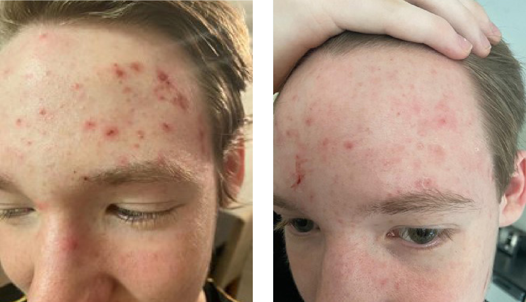
The patient's teenage son (patient three) was also struggling with skin issues and was unable to control his acne with antibiotics and the usual astringent cream/washes. The author allocated him 2 weeks of 15 minutes 415nm exposure twice a week.
Reviewing the patients
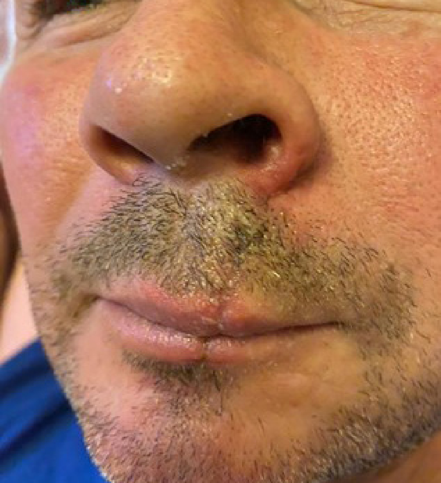
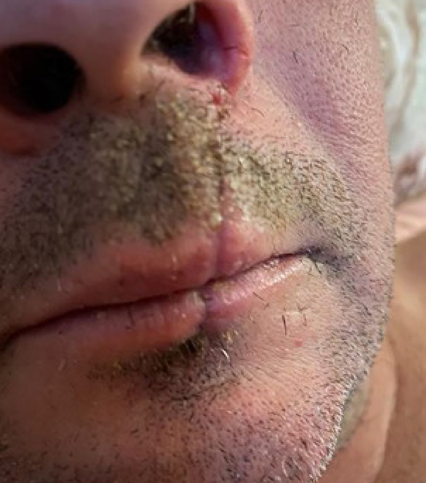
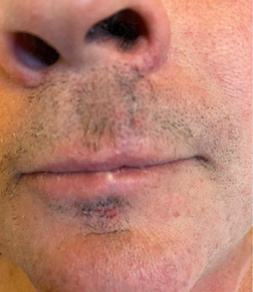
The author agreed to review patient 1 at the end of the 6-week period to reassess the next step. Pictures of his healing progress were sent at regular intervals. The first photograph (Figure 2) was sent on 25 January 2021 (15 days post-accident) and showed that healing was progressing well, and his comfort level was better. However, he was still having to drink through a straw due to a lack of movement. On 31 January 2021, there was a steady ongoing improvement, although the mid-lip section had started to pucker a little and subcision seemed likely with a possible need for dermal filler at a later date (Figure 3).
On 11 February 2021, patient 2 sent photographs of her son (patient 3) after six treatments. The images showed that all inflammation was reduced and there was also a reduction in pustules (Figure 4). The following day, patient 2 also sent pre- and post-treatment photographs of her rash following the treatments (Figures 5–7), as well as a note:
‘The difference is really impressive, isn't it? Honestly, I've had that rash for about 4 months and no creams worked at all. The improvement was after the first treatment and it just got better and better’
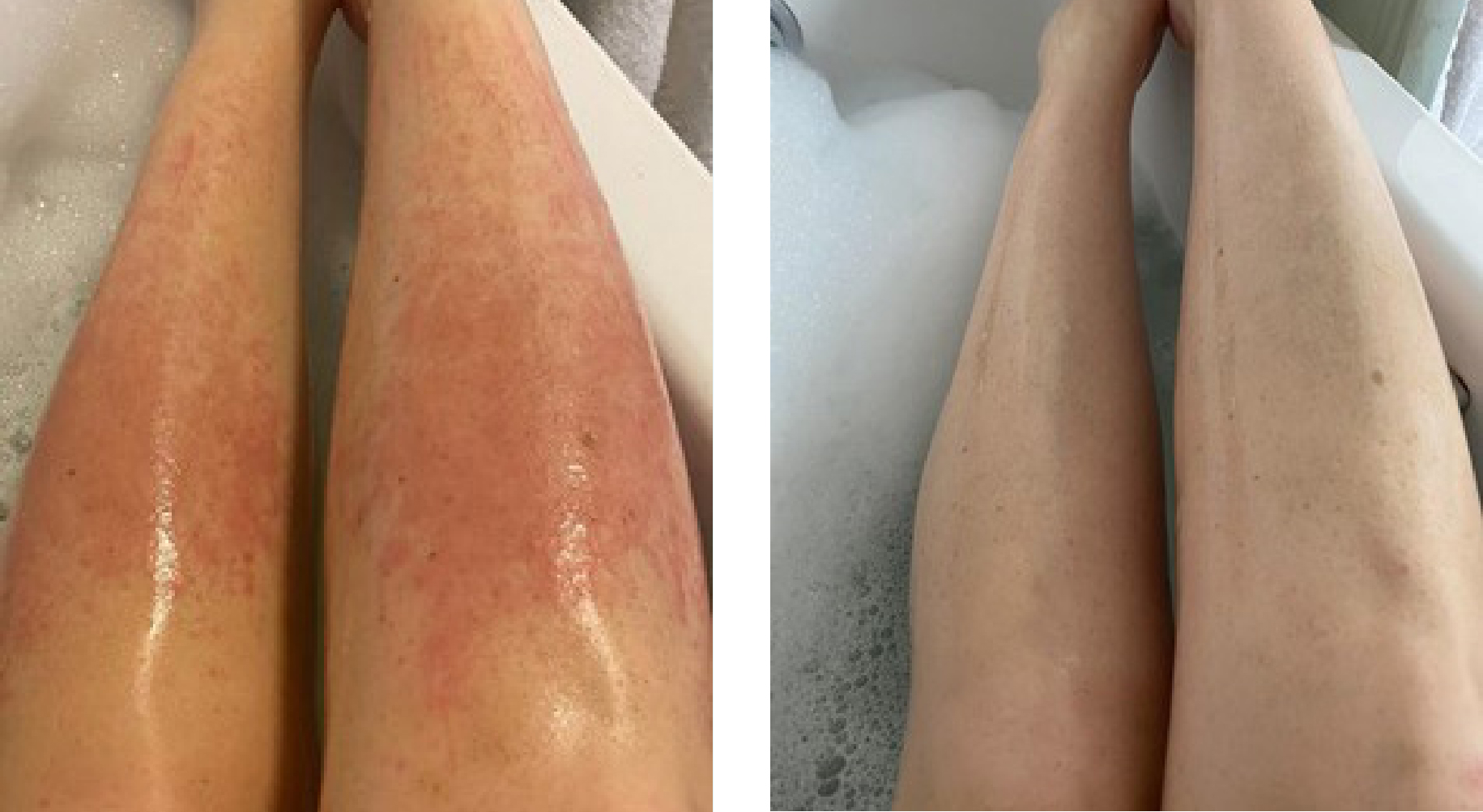
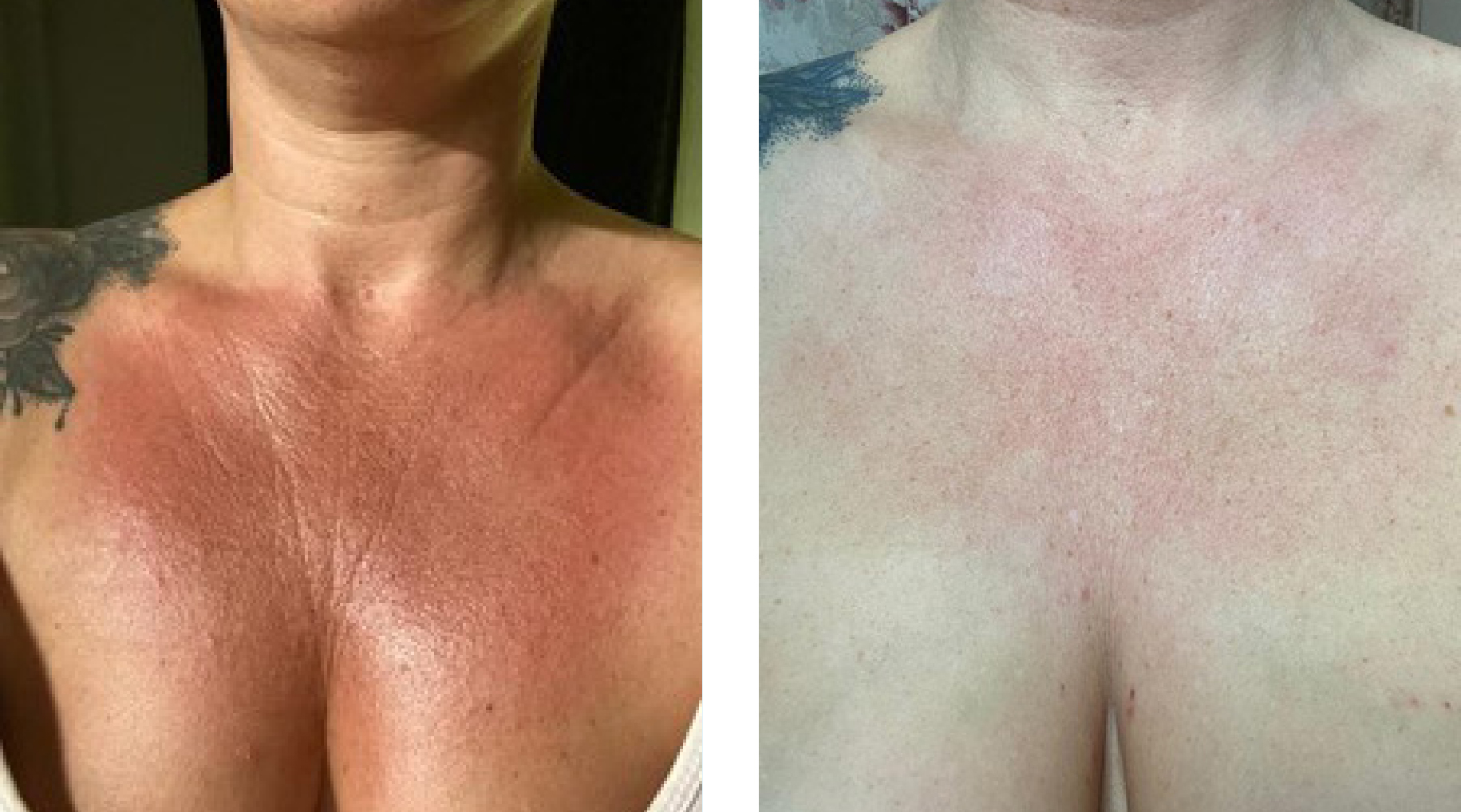
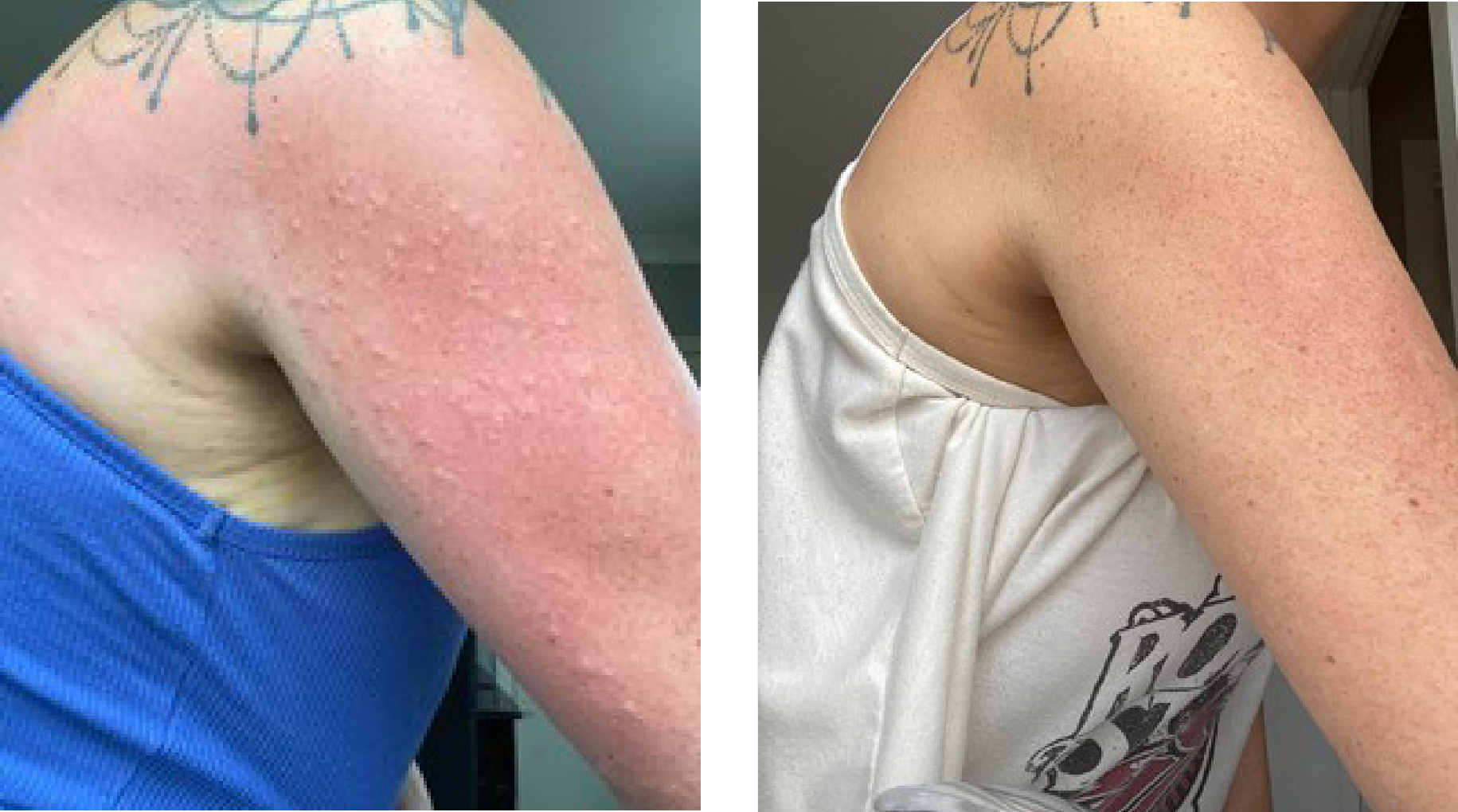
The next photographs of patient 1 arrived on 22 February 2021. All signs of inflammation were gone, and the scar was uniform and neater than it was previously. His partner (patient 2) had been discharged by her dermatologist following a review, and no medications were required to be used. There has been no return of symptoms since completing her sessions.
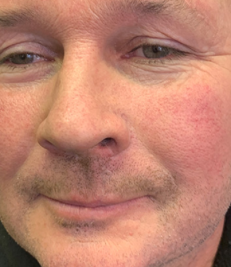
Follow-up pictures were received on 16 March 2021 (Figure 8) (10 weeks since the accident), and patient 1 reported that he was able to drink without a straw, as the scar is more mobile.
He was reviewed on 8 April 2021, and, due to the healing progress, the author decided to introduce needling (0.3mm needle depth pen device) to assist with collagen growth (Figure 9). One session per week for 6 weeks with 0.8% HA applied was carried out to maintain hydration. The rest of the year continued, and he continued to report positive healing.
Healing progression
COVID-19 restrictions limited non-essential reviews, so, when they eased, the author was able to carry out a review in their clinic, which showed that the scar was continuing to heal well. The author decided to let time progress and review at the anniversary of the injury to ascertain whether platelet-rich plasma (PRP) may be needed to assist with the scarring to the actual lip, which caused a little uneven texture.
On 19th January 2022, patient 1 came for his official review (Figure 10) and kindly provided a video testimonial for the company's medical aesthetic arm launch www.silvery.blue. The final picture speaks volumes.
The crux of the case study is to demonstrate how versatile medical aesthetics practice can be, the skills that can be used and how having a solid foundation of knowledge of how products work and synergise can benefit a variety of patients with short-term issues and long-term goals.