References

Berk DR, Bayliss SJ. Milia: a review and classification. J Am Acad Dermatol.. 2008; 59:(6)1050-1063 https://doi.org/10.1016/j.jaad.2008.07.034

British Association of Dermatologists. Sunscreen fact sheet. 2013. http://www.bad.org.uk/skin-cancer/sunscreen-fact-sheet (accessed 2 February 2021)

Milium cysts in adults and babies. 2018. http://www.healthline.com/health/milia (accessed 4 February 2021)

Milia causes and treatment options. 2021. https://www.harpersbazaar.com/uk/beauty/skincare/a18542004/milia-causes-and-treatments (accessed 8 January 2021)

MediNiche. Caring for the skin around the eyes. 2021. http://www.mediniche.com/ocularskincare.html (accessed 11 February 2021)

Anatomy of the skin. 2021. http://www.robertmillermd.com/anatomy_of_the_skin.html (accessed 2 February 2021)

Morris G. Skin blemish removal with diathermy: a practical guide to Levels 4 and 5.: Sterex; 2018

What is the stratum corneum?. 2019. http://www.healthline.com/health/stratum-corneum (accessed 8 January 2021)

Non-surgical removal of milia: treatment and aftercare

02 March 2021
Volume 10 · Issue 2

Abstract

Although harmless, milia—small, white spots most commonly found near the eyes—are a cosmetic concern and must be removed safely by a trained professional. Gillian Huntley details the types of milia and explains how they are treated

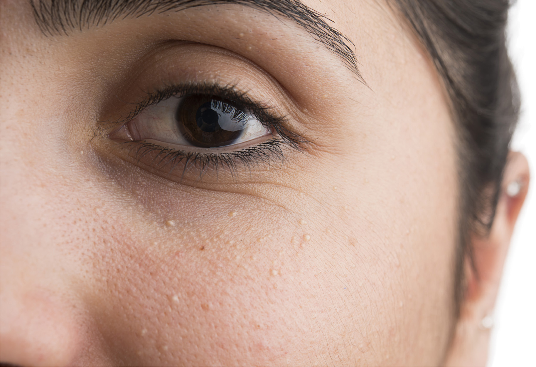
Milia come in a variety of different sizes and can affect any Fitzpatrick skin type and people of all ages

Milia are small, round milky white spots that can vary in size up to 1–2 mm. They are also often called whiteheads or milium plugs and are keratin-filled cysts that are harmless and most commonly found in dry skin, on the face but mainly in and around the hollow orbit of the eye, tear trough and the infraorbital area (Kinman, 2018). They are located under the epidermis and within the dermis, so they cannot be squeezed or rubbed away.

Arising from an undeveloped sebaceous gland or sweat gland they are formed within the dermis where these glands are found and become more visible as they migrate up to the epidermis. (Morris, 2018)

The anatomy of the skin around the eye area is called the adnexa (MediNiche, 2021).

Eyelid skin has several layers: the basal layer is the deepest and continually reproduces new skin cells, which then migrate to the surface where they become dry and flat. They are then exfoliated and flake off in a process called desquamation. As the skin ages, this process takes longer, from around 30 days in earlier ages to 37 days once aged over 50 years. Due to the skin build up, and as the eye area does not contain oil sweat glands, it is one of the driest areas of the skin on the face and body (Sawyers, 2019).

Skin is thickest on the palms and the soles of one's feet, around 1.5 mm compared to the eyelid orbit area, which is only 0.05 mm thick (Miller, 2021). As the body ages, the skin loses elasticity and muscle strength, and the levitator muscles of the eye become weaker. Consequently, this can cause a drooping or double-looking eyelid. It is in these creases that oil and sebum can get trapped and cause milia to appear.

As with many skin conditions, milia come in a variety of different sizes and can affect any Fitzpatrick skin type and all ages. Congenital milia can occur in newborns, as well as adults, although the exact causes are unknown (Berk and Bayliss, 2008). However, it is known that it more frequently occurs in adults with dry, dehydrated skin or as a build-up of excess skin cells that trap sebum. It can also be caused by the range of skincare products used, including using sun lotions that are too ingredient-rich for this delicate area of the body (British Association of Dermatologists, 2013).

Types of milia

Primary milia

These can develop in childhood, adolescence and in adults of both sexes and are commonly seen in multiple eruptions around the eyelids, cheeks, forehead and nose but can also appear elsewhere on the body. They are rarely associated with hereditary factors (Morris, 2018).

Secondary milia

Secondary milia can arise from the site of an injury, damaged eccrine glands or blistering of epidermal tissue (Morris, 2018).

The sweat ducts become blocked as a result of using oil-based products around the thin skin of the eye, and these beauty products, together with a lack of sleep, poor diet, poor hygiene, poor cleaning, smoking or steroids, can cause the milia (March and Schroeder, 2021).

Pre-treatment of milia

A pre-treatment record card and photos should be filled out with the patient prior to any treatment.

Although there is no certainty as to the exact causes, it is important to look at some of the possible associated causes with each patient during the consultation process by carrying out a full skin evaluation to classify skin type and hydration levels.

When treating patients, it is essential that a detailed medical questionnaire is completed to find out how long the patient has had the milia, which skincare products they use, their face washing and cleansing process, how often they wash the area, which products are used for cleansing (if any) and whether they use wipes, cleansers or eye make-up removers. It may be possible that the relationship between the products used on the affected areas may be playing a key part in the occurrence of milia.

Through this process, other potential contributing factors can be identified.

Although there is debate among professionals as to whether there is any correlation between the occurrence of milia and a high cholesterol diet, at this stage, it is still important to take diet in consideration.

Medical contraindications

Before the practitioner proceeds with the treatment, there are strict guidelines regarding any pre-procedure contraindications that could prevent the removal from going ahead. As mentioned, a medical questionnaire must be filled out by the patient prior to treatment and, should there be any concerns regarding a health condition, then a doctor's permission must be gained, although it is uncommon. Those with major health conditions, such as heart problems or diabetes, or patients who take warfarin, are pregnant, have keloid scarring, haemophilia or a pacemaker will need to seek approval from a doctor to ensure that they are a safe candidate for the treatment.

Patient questions

The author recommends asking the following questions before milia treatment:

  • How long ago did you notice the milia?
  • Have you tried to remove the milia yourself?
  • Do you suffer from dry skin?
  • Do you take any supplements?
  • What products do you use on your face?
  • How much water do you drink daily?

Patient reassurance is important during the pre-treatment assessment. The eye area is an extremely delicate area to treat, as the skin is very thin, and, because of this close proximity to the eye, the patient can become nervous, pull away, blink a lot or their eyes can water. Establishing patient trust and understanding prior to treatment will help reduce these natural reactions during the treatment process (Figure 1).

Figure 1. One patient during the milia treatment process

Treatment

It is recommended to photograph the area before and after the treatment (Figure 2).

Figure 2. Before and immediately after milia treatment

There are two methods of milia removal that the author uses. First, the size and position of each lesion is assessed, and the options to remove are:

  • For large milia: this can be extracted using a sterile lancet and incising the area, creating a ‘hot cross bun’ technique that allows the hard lump to simply be expelled. Although the treatment is not painful, it can cause some clients a little discomfort with a pricking sensation as each milia is targeted. Slight scabbing will also occur following treatment as the skin heals
  • For smaller milia: the second optional removal treatment is with using a diathermy machine and a sterile single-use probe. A new probe (sealed in an individual pouch and gamma-irradiated) is inserted into each milium, instantly dissolving the fatty tissue via heat cauterisation. This is inserted into each blemish at a 90-degree angle to dissolve the core, and the low intensity current will work with minimal discomfort to the patient, although the sensation felt by individuals does vary (Morris, 2018).

This advanced cosmetic procedure allows for an easy removal of any milia and has minimal side effects or scarring. Advanced electrolysis involves sending a high frequency alternating current through a selected size of needle to destroy the targeted cells to remove the blemish. The diathermy heat will desiccate the fatty ball. The probe has to be inserted through the skin to a depth of approximately less than 0.5 mm. Some blood can appear at this stage, but the area will quickly dry up. During this process, the treatment area is gently and carefully wiped with saline, and once treatment has been completed, a gel or thick non-oily product is used to seal the openings and protect from infection. The area will have tiny red dots post-treatment but, in a healthy patient, these will disappear fairly quickly.

Post-treatment

Immediately after treatment, erythema or oedema may be experienced in the area. After a few days, the area will appear to have small scabbing where the treatment was carried out.

A soothing aloe vera or cooling gel or petroleum jelly should be applied immediately after the treatment to cool and soothe the area. It is vital that the product does not go into the actual eye to protect the delicate eye tissue, as it is possible that it could cause stinging or irritation.

Following treatment, the patient needs to avoid direct sunlight and getting the area wet, nor should they use steam rooms or saunas or go swimming. Therefore, this treatment is not recommended prior to any sun trips or holidays.

It is recommended that the treated area is kept free from make up during the healing process, which can take up to 1 month providing the correct after care instructions are followed. Pre-arranged special occasions may affect the patient's ability undergo treatment.

The patient should be advised that the area treated will form tiny scabs that need to heal naturally. They must avoid rubbing or knocking the area, as the scabs could accidently be removed and cause potential scarring of the skin.

On skin where there is a highly concentrated area of milia, treatments will need to be spaced out over a period of time to allow for healing between sessions and to avoid overtreating areas. There is also the possibility that larger individual milia may need more than one session to break down the cells before they disappear. Due to the fineness of the eye area skin, bruising can occur.

The possibility of recurrence

Once the treatment has finished, it is important that the patient adheres to the practitioner's aftercare instructions to ensure that the most effective healing can take place. The author uses a written aftercare sheet and a specific aftercare product to give to her patients. If the patient continues with incorrect products around the eye, then the milia could possibly return.

Advice for avoiding milia

  • Double cleanse the eye area daily. First, with a non-oily eye makeup remover and, second, with a cream cleanser, making sure that all traces of eye makeup product are removed using a soft damp cotton pad
  • Avoid using heavy oil products around the eye area
  • Avoid sun exposure to the face and always wear an SPF 50 on the face, while checking that the products contain no oil
  • Assessing diet and lifestyle: in particular, more sleep and reduced cholesterol-rich foods (for example, eggs and meats)
  • Taking vitamins A, B and D supplements may help to reduce milia
  • Always wear good quality sunglasses.