References

Jagus D, Skrzypek E, Migds B, Wozniak W, Mlosek RK. Usefulness of Doppler sonography in aesthetic medicine. J Ultrason. 2020; 20:(83)e268-272 https://doi.org/10.15557%2FJoU.2020.0047

Mlosek RK, Migda B, Skrzypek E, Sloboda K, Migda M. The use of high-frequency ultrasonography for the diagnosis of palable nodules after administration of dermal fillers. J Ultrason. 2020; 20:(83)e248-253 https://doi.org/10.15557/jou.2020.0044

Qiao j, Jia QN, Jin HZ Long-term follow-up and diffusion pattern of hyaluronic acid in nasolabial fold correction through high-frequency ultrasound. Plast Reconstr Surg. 2019; 144:189-196e https://doi.org/10.1097/prs.0000000000005848

Schelke LW, Decates T, Kadouch J, Velthuis P. Incidence of vascular obstruction after filler injections. Aesthet Surg J. 2020; 40:(8)NP457-NP460 https://doi.org/10.1093/asj/sjaa086

Schelke LW, Velthuis P, Kadouch J, Swift A. Early ultrasound for diagnosis and treatment of vascular events with hyaluronic acid fillers. JAAD. 2019; S0190–9622:(19)32392-32398 https://doi.org/10.1016/j.jaad.2019.07.032

Scotto di Santolo M, Massimo C, Tortora G Clinical value of high-resolution (5-17 MHz) echo-color doppler (ECD) for identifying filling materials and assessment of damage or complications in aesthetic medicine/surgery. Radiol Med. 2019; 124:(6)568-574 https://doi.org/10.1007/s11547-018-0969-1

Use of duplex ultrasound in facial aesthetics. 2021. http://www.prime-journal.com/use-of-duplex-ultrasound-in-facial-aesthetics (assessed 15 December 2021)

Wortsman X, Wortsman J. Sonographic outcomes of cosmetic procedures. AJR Am J Roentgenol. 2011; 197:(5)W910-918 https://doi.org/10.2214/ajr.11.6719

Ultrasound imaging in aesthetic practice and what to consider when thinking about its adoption
02 April 2022
Volume 11 · Issue 3

Abstract

In the first part of this series, Amy Miller explores the considerations that aesthetic clinicians should make when implementing ultrasound imaging in their practice

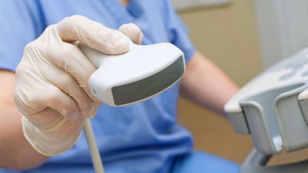
Facial ultrasound imaging has become an increasingly popular topic in the field of medical aesthetics. A common tool in almost every other area of medicine, it has been slow to penetrate the field of aesthetics. Over 10 years ago, Wortsman and Wortsman (2011) published an article on sonographic outcomes of cosmetic procedures, but ultrasound imaging is only now gaining traction in this field. This is partly because older ultrasound machines tended to have poor resolution of the tiny, superficial features of the face and could be prohibitively expensive. However, more recently, better quality and less expensive ultrasound machines have been mass marketed to the medical aesthetics sector, making it easier for practitioners to adopt this technology. However, questions remain: is it truly revolutionary for aesthetics, and is it really worth it?

Potential barriers to ultrasound use

Despite improvements in the technology and reductions in cost, there are still significant barriers to the ubiquitous use of ultrasound in medical aesthetics. It has a significant learning curve that requires time and dedication on the part of the user. Remember that there are professionals who dedicate their entire career to ultrasound imagery. Ultrasound effectiveness is operator-dependent, and it is a skill that should be practised regularly to ensure that the practitioner remains well-versed in its use. Even though the equipment has become more affordable, the training and education on the use of the machine can add up quickly. Time and money are not resources to be spent frivolously, so it is essential that practitioners know how the advantages and disadvantages of ultrasound adoption weigh out for them and their practice.

Benefits of ultrasound

There are many benefits of ultrasound use, and these include: the ability to visualise all facial anatomical layers in real time, which is helpful for reinforcing the user's 3D anatomical knowledge base; the ability to check for aberrant, unusual or simply non-textbook anatomical architecture; the ability to locate and measure the depth of vessels to avoid intravascular injections; guided dermal filler injections and guided therapeutic injections, such as hyaluronidase (Weiner, 2021); evaluation of previously placed fillers and discovery of evidence on their composition, age, depth and spread, etc (Qiao et al, 2019; Scotto di Santolo et al, 2019); evaluation of filler complications, including nodules, delayed onset nodules, biofilms and granulomas, etc, and distinguishing these from bulky filler deposits (Mlosek et al, 2020); and the ability to locate vascular occlusions or stenosis (Jagus et al, 2020) for targeted, efficient and immediate treatment to avoid a CVA or retinal injuries/blindness.

Other, less tangible, benefits of ultrasound use include the injector's peace of mind and increased confidence while administering treatments. The patient may also have increased confidence in their injector and feel less stressed during injections. Patients may be more apt to return to the same injector, especially if they have the perception that a practitioner is more familiar with their individual facial anatomy. The possibility of repeat business is a reason that a practice administrator may be persuaded to spend money on equipment and training.

The incident that led to the author being pro-ultrasound was not, as one might suspect, due to the enhanced safety profile of this technology (she felt that she had been injecting safely for 20 years). It was the ability to treat a tear trough protrusion of hyaluronic acid (HA) dermal filler of several years' duration (Figure 1). Several ‘blind’ attempts at dissolving the pocket of filler with hyaluronidase resulted in shrinkage, but not elimination, of the pocket and damage to the surrounding supportive tissue. The ability to see the needle penetrate the HA pocket (Figure 2) gave the author the confidence boost needed to inject a high enough dose of hyaluronidase to eliminate the pocket (Figures 3 and 4). Now, the patient no longer has to use elaborate makeup camouflage techniques and can pose for photographs without hiding the area with her hair.

Figure 1. Patient prior to treatment with visible hyaluronic acid nodule
Figure 2. Ultrasound picture of nodule, notice the posterior enhancement indicating the sound waves are travelling right through the nodule, indicating a liquid or aqueous gel substance
Figure 3. The patient 3 weeks following treatment
Figure 4. Ultrasound image of the area, indicating resolution of the hyaluronic acid pocket

Protocol for use

If a practitioner does decide to implement ultrasound imaging in their clinic, a protocol for use needs to be established. Will every patient be scanned and have vascular mapping? Will only the areas of injections be imaged? Will only high-risk injection areas be imaged (for example, the nose, glabella and temple)? Will the device only be used for evaluation of oedema, nodules and other side effects, or will it be reserved for emergency use? Regardless of the chosen protocol, standard and consistent documentation is a must.

As the field of aesthetic medicine grows, complications from dermal filler injections also continue to grow. In 2011, the Eramus Medical Centre in Rotterdam established an outpatient clinic to deal with dermal filler adverse events, whether they are patient-related (such as allergic reactions), product-related (for example, inflammatory responses) or injector-related (for example, misplacement or overcorrection) (Schelke et al, 2019). Schelke et al (2019) estimated that they saw complications of intravascular injection of fillers, including necrosis and blindness, approximately twice per month. In a follow-up report, Schelke et al (2020) attempted to quantify the overall rate of vascular adverse events (VAEs), placing it at 0.015% chance per treatment or one in 6558 treatments, but noting that this may be an underestimation due to lack of recognition and underreporting (Schelke et al, 2020). They stated: ‘With a risk of 1:6800 treatments, many physicians will encounter this event more than once in their career’.

Conclusion

From a safety standpoint, is it worth the expense, training and time required to be prepared for an event that may or may not happen once or twice in someone's career? The true issue may not be the overall rate of VAEs, but the devastating consequences of a VAE to a patient. Blindness or stroke should never happen as a result of an aesthetic procedure, especially now that tools are available to help prevent and reverse filler occlusions. If the incidence of these severe injuries continues to increase, the sector needs to be ready for possible political and Government involvement, and new regulations coming from outside the health sector. Additionally, consider the possibility that future practitioners may think that it was ludicrous to inject dermal filler without imaging assistance, and that, eventually, ultrasound will most likely become the standard of care. In the author's opinion, it is simply a matter of time before it is common to be found in medical aesthetic clinics. So, it is not a matter of if, but when, to learn how to use facial ultrasound imaging.

Ultrasound imaging is a tool that has finally evolved to find a place in cosmetic injectable practice. Despite the time and resources required to be efficient with ultrasound imaging, it is a tool that can increase the safety of injectable filler and aid in the diagnosis and treatment of adverse events, vascular or otherwise. If VAEs continue to increase, the public will demand changes and the medical aesthetics sector may find itself regulated by non-medical lay people. Patient safety, injector peace of mind and the ability to diagnosis and treat complications are all compelling reasons to become proficient with ultrasound use.